Academic Staff
Chair of Radiology and Diagnostic Imaging Department
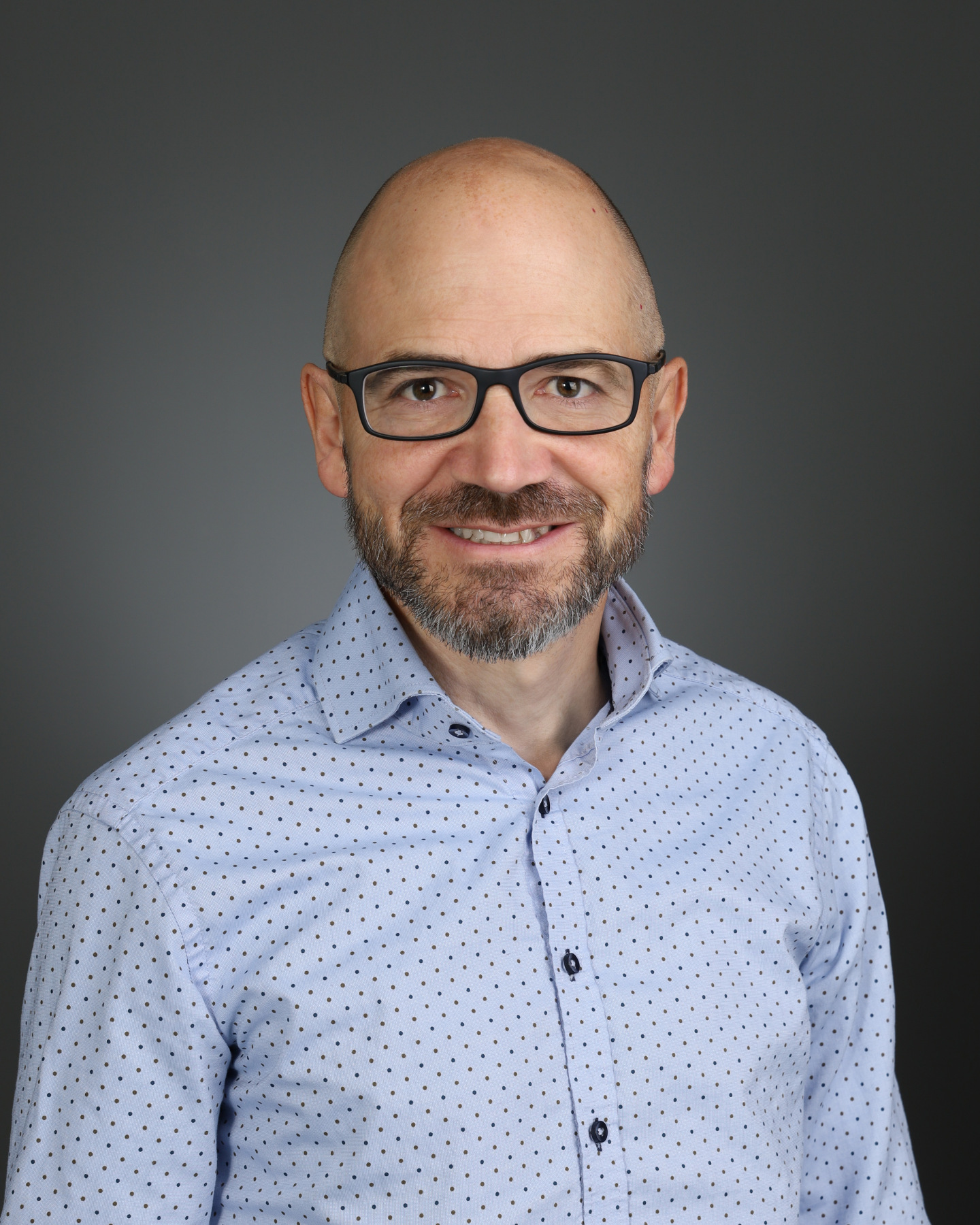
Dr. Derek Emery, Professor
Contact - Publications
Dr. Derek Emery is the appointed Chair of Radiology and Diagnostic Imaging, partner with Medical Imaging Consultants (MIC) and the Medical Director of the Peter S. Allen in vivo MR Research Facility. He has clinical fellowships in Neuroradiology from the University of Toronto and Interventional Neuroradiology from Baptist Memorial Hospital, Memphis. Dr. Emery current research focuses on appropriateness and utilization of imaging.
Geographical Full- Time (GFT)
Dr. Jonathan Abele, MD
Contact - Publications
Dr. Jonathan Abele is an Associate Professor in the Radiology and Diagnostic Imaging Department at the University of Alberta and is a partner with Medical Imaging Consultants (MIC). He has a Bachelors of Science (BSc.) from the University of Alberta (1995), a MD from the University of Western Ontario (1999), and completed residency training at the University of Alberta in Diagnostic Radiology (2004) and Nuclear Medicine (2006). Dr. Abele's academic interests include the topics of hybrid imaging, SPECT/CT, PET/CT, and the clinical implementation of new diagnostic radiotracers. He is the co-director of the Canadian Nuclear Medicine resident review course and has been an active board member with the Canadian Association of Nuclear Medicine.
Dr. Christian Beaulieu, PhD
Contact - Website - Publications
Dr. Christian Beaulieu is a Professor (cross-appointed with Department of Biomedical Engineering), Canada Research Chair (Tier 1) on MRI on Brain Micro-structure, Scientific Director of the Peter S. Allen Magnetic Resonance Imaging (MRI) Research Centre, and University of Alberta Canadian Institutes of Health Research (CIHR) University Delegate. His lab focuses on the technical development of novel MRI methods as well as their application to a number of clinical disorders. The overarching goal is to drive the next generation of MRI which can detect and quantify micro-structural features in brain (and other tissues) that cannot be measured with standard MRI. Working with clinical researchers, the MRI innovations are used currently to discover new information on people with stroke, epilepsy, multiple sclerosis, neurodevelopment disorders, typical development and aging over the human lifespan, and prostate cancer. MRI student (MSc/PhD/PDF) projects are suitable for multi-disciplinary backgrounds including undergraduate degrees in engineering, physics, biology, psychology, medicine, neuroscience, computing science, physiology, etc. The MRI projects can be on developing and/or optimizing new pulse sequences and methods, image processing and analysis, hardware design and construction, and/or human studies using existing methods/protocols. See Google Scholar for a full list of publication to see the scope of research.
Dr. Christopher Fung, MD, FRCPC, DABSR, FSRU
Contact - Publications
Dr. Christopher Fung is an Associate Professor in the Radiology and Diagnostic Imaging Department at the University of Alberta. His academic interests have focuses on guideline development and review, with a particular focus on abdominal imaging. Dr. Fung has assisted with the development and publication of both natural and international guidelines on renal lesions, pancreatic cysts, gallbladder polyps, heptocellular carcinoma surveillance, and hepatobiliary incidental findings. He has also been involved in research regarding prostate cancer from an access perspective and best imaging/interventional practice. Dr. Fung has specific interest in ultrasound and development/evaluation of novel sonographic technology such as micro-ultrasound in prostate cancer and elastography of liver disease.
Dr. Abhilash R. Hareendranathan, PhD
Contact - Website - Publications
Dr. Abhilash Rakkunedeth Hareendranathan is an Assistant Professor in the Department of Radiology and Diagnostic Imaging at the University of Alberta, specializing in artificial intelligence (AI), medical image analysis, and ultrasound imaging. With extensive international research experience, he has collaborated with multidisciplinary teams across Singapore, Germany and Canada.
Dr. Hareendranathan earned his PhD. from Nanyang Technological University (NTU) in Singapore, where hsi research focused on developing machine learned techniques for movement compensation in image-guided, non-invasive surgical procedures. Post-PhD he joined the ultrasound team at Panasonic R&D Center in Singapore and developed novel ultrasound beamforming approaches for carotid imaging. Subsequently, at Curefab in Germany, he worked as an image segmentation specialist.
Dr. Hareendranathan served as the Research and Development Leads at Medo.ai, an ultrasound AI-focused start-up based in Singapore and Edmonton. His team developed innovative AI-driven solution for medical ultrasound.
Dr. Hareendranathan is currently a Principal Investigator at the Northern Institute for Deep Leaning in Ultrasound (NIDUS). His research focuses on developing AI approaches for automatic biomarker discovery, improving assessment reliability and efficiency, and optimizing clinical workflows to empower minimally trained operators. His work aims to bridge AI advancements with clinical needs to enhance patient outcomes.
Dr. Tracey Hillier, BScN, MD, CCFP, FRCPC, MEd
Contact
Dr. Tracey Hillier is the Executive Dean and Director of the Alberta Institute (an international medical school collaboration between the University of Alberta and Wenzhou Medical University in China); and an Associate Professor in the Department of Radiology and Diagnostic Imaging at the University of Alberta. Prior leadership as Associate Dean MD Program, University of Alberta. Completed residencies in Family Medicine (McMaster University), Radiology (University of Alberta) and Emergency Trauma Radiology Fellowship (University of British Columbia). Clinically, focused on Breast Imaging, academically focused on EDI and International Medical Education.
Dr. Hillier is a member of the Mi'kmaq Qalipu First Nation. Co-director of the Situated Knowledges: Indigenous Peoples and Places University of Alberta, a member of the Vice-Provost Indigenous Programming and Research Advisory Council, the Indigenous Research Strategy Task Force, the Faculty of Medicine and Dentistry EDI and Indigenous Initiatives Multi-Institute Committee and the Steering Committee for Indigenous Initiatives and Equity, Diversity and Inclusion in Clinical Trials. Prior service on the University of Alberta Vice-President Research and Innovation Advisory Committee and the Intellectual Property & Commercialization Policy and Process Review Committee. A member of the Indigenous Physicians Association of Canada.
A CIHR Accelerating Clinical Trials Consortium Indigenous Subcommittee and ACT training committee member. Member of the Board of Directors for the Canadian Emergency Trauma and Acute Care Radiology Society and prior Vice-Chair for the Canadian Association of Radiologist Equity, Diversity and Inclusion Working Group.
Dr. Jacob Jaremko, MD, PhD, FRCPC
Contact - Website - Publications
Dr.Jacob Jaremko is a Professor of Radiology and Adjunct Professor of Computing Science at the University of Alberta, a practicing board-certified Pediatric and Musculoskeletal radiologist and partner of Medical Imaging Consultants, and co-founder of two startup companies including Medo.ai. He has a PhD in Biomedical Engineering. Jaremko is a Canada CIFAR AI Chair and Fellow of the Alberta Machine Intelligence Institute. His research has focused on developing objective imaging biomarkers of disease in ultrasound and MRI, and on implementing AI-augmented medical imaging diagnostic tools at the clinical point of care -- building the 21st century stethoscope.
Dr. Alexander Kwan, PhD
Contact - Publications
Dr. Alexander Kwan is an Assistant Clinical Professor in the Radiology and Diagnostic Imaging Department at the University of Alberta. He is a diagnostic medical physicist working for Alberta Health Services since 2007. Dr. Kwan received his PhD from the University of Alberta, postdoctoral training at University of California, Davis and is certified in diagnostic medical physics by the American Board of Radiology. His primary research interest is in dedicated breast CT imaging.
Dr. Robert G. W. Lambert, MB BCh, FRCR, FRCPC
Contact - Publications
Dr. Lambert graduated from Queen's University, Belfast, Northern Ireland in 1980, did radiology residency in Belfast and a two-year MSK radiology fellowship in Toronto,Ontario. Dr. Lambert was head of MSK radiology at the University of Alberta from 1982-2002 and was at the forefront of MSK radiology innovation introducing many MSK MRI and interventional MSK services to Canada. Dr. Lambert was Chair of the Department of Radiology from 2002-2016. He was awarded the Fellowship of the Canadian Association of Radiologists in 2021. Dr. Lambert is a founding member of the Canadian Society of Skeletal Radiology (CSSR) and a member of BSSR, ESSR, SSR, ISS, CAR, RSNA, ASAS and SPARTAN.
Dr. Lambert's primary research interests are MSK MRI and imaging of arthritis. This incuse the development and clinical validation of image-based definitions and scoring tools for assessment of spondyloarthritis (SpA) and osteoarthritis, and randomized trials of arthritis therapy. He has published over 170 peer-reviewed articles, 5 book chapters, and hundreds of abstracts. Among his most notable innovations are the development of the SPARCC MRI scoring systems for inflammation and structural damage in the sacroiliac joints and spine that are now industry standard in SpA clinical trails.
Dr. Lawrence H. Le, PhD, MBA
Contact - Publications
Dr. Le received his PhD in physics and MBA in finance and technology
commercialization from the University of Alberta, Edmonton, Canada. He held a Natural
Sciences and Engineering Research Council of Canada (NSERC) Postdoctoral
Fellowship at Schlumberger-Doll Research, Ridgefield, CT, USA. He started his medical
physics residency training at the Department of Radiology and Diagnostic Imaging
(DRDI), University of Alberta, in 1994. He is currently a Clinical Professor leading the
Graduate Program at DRDI and a Senior Medical Physicist with Alberta Health
Services, Edmonton. He is also an adjunct professor in Fudan University, China. His
research interests include ultrasound imaging, signal and image processing, wave
propagation modeling and inversion, and machine learning.
Dr. Gavin Low, MBChB, MPhil, MRCS, FRCR
Contact - Publications
Dr. Gavin Low is Professor in the Radiology and Diagnostic Imaging Department at the University of Alberta and is a partner with Medical Imaging Consultants (MIC). He was educated at the University of Edinburgh (MBChB), University of Cambridge (MPhil), underwent clinical training in radiology in Glasgow, Scotland and completed a body imaging fellowship at the University of Alberta. Dr. Low's main research interests are in transplant imaging (liver, renal, pancreas and islet), in imaging characterization of focal lesions in the abdomen using ultrasound, CT, MRI and magnetic resonance elastography.
Dr. Michelle Noga, MD, FRCP
Contact - Website - Publications
Dr. Michelle Noga is a pediatric cardiac radiologist with an interest in 3D display/technologies, machine leaning applications in postprocessing and image segmentation, and 3D ultrasound. She has a significant interest in interdisciplinary research and is director of the Servier Virtual Cardiac Centre and the Alberta Health Services Medical Imaging Consultants Research Chair. Dr. Noga is a partner of Medical Imaging Consultants and vice-char of the Pediatric Radiology exam board, Royal Collage of Physicians and Surgeons of Canada. She has authored over 115 publications on multiple subjects, predominately in pediatric radiology, machine learning, 3D display and cardiac imaging. Dr. Noga enjoys mentoring and teaching students and residents of all levels in clinical medicine and research.
Dr. Kumar Punithakumar, PhD
Contact - Website - Publications
Dr. Kumar Punitakumar is an associate professor in the Department of Radiology and Diagnostic Imaging at the University of Alberta, where he holds the Alberta Health Services Chair in Diagnostic Imaging. Additionally, he serves as the Operational and Computational Director of the Servier Virtual Cardiac Centre at the Mazankowsku Alberta Health Institute. He earned his PhD in Electrical and Computer Engineering from McMaster University and worked as research scientist for four years at GE Healthcare. Since joining the University of Alberta in 2012, he has supervised over 70 highly qualified personnel, including postdoctoral fellows and PhD, MSc, and undergraduate students. His research portfolio comprises more than 120 peer-reviewed publications and nine patent applications.
Dr. Punithakumar received the Industrial Research and Development Fellowship from the National Sciences and Engineering Research Council of Canada in 2008 and the Outstanding Associate Editor award from IEEE Access in 2018 and annually from 2020 to 2023. His research interests include medical imaging analysis and visualization, machine learning, computer vision, robotics, information fusion and object tracking.
Dr. Richard Thompson, PhD
Contact - Publications
Dr. Richard Thompson is a Professor of Radiology and Diagnostic Imaging at the University of Alberta. His research focus is on development and application of structural (fibrosis, edema, inflammation) and functional MRI with a focus on the heart, lungs, skeletal muscle liver and fat with an emphasis on quantitative imaging imaging with exercise challenge. Key areas of application include heart failure, cardio-oncology and COVID-19 with emphasis on exercise capacity and metabolism.
Dr. Alan Wilman, PhD
Contact - Website - Publications
Dr. Alan Wilman's research is in the MRI of the brain with a focus on developing and applying new methods of MRI acquisition or image processing to enable measurement of quantitative relaxation and magnetic susceptibility. It is the application of the developed methods to neurological diseases with an emphasis on multiple sclerosis. The training environment is the Peter S. Allen MRI Research Centre, offering world-class research MRI equipment within our hospital environment. Dr. Wilman's training is in MRI physics and graduate students that usually have a technical expertise with engineering or physics background. To better understand his research, see PubMed.
Faculty Service Officers (FSO)
Dr. Karim Damji, B.Sc, E.E, M.Sc., P. Eng.
Contact - Publications
Al-Karim Damji is a Faculty Service Officer in the Department of Radiology and
Diagnostic Imaging at the University of Alberta, where he is responsible for the
maintenance of the Siemens 3T Prisma scanner and the 4.7T MRI scanner at the Peter
S. Allen MR Research Centre. He also maintains the Department of Oncology Siemens
mMR 3T PET-MR scanner, and the Department of Pediatrics Bruker 3T Preclinical
PET-MR scanner. He earned his M.Sc in Applied Sciences in Medicine at the University
of Alberta. He’s also completed training for servicing several MR and PET scanners
(Siemens’ Symphony/Harmony MRI, MRI Magnets and 4K Cryocoolers, Aera/Skyra
MRI, and PET Basics courses), and mMR PET-MR Gantry courses). He also develops
MRI hardware, especially RF coils and other required hardware to support specific MRI
research projects.
Dr. Rob Stobbe, PhD
Contact - Publications
Dr. Rob Stobbe is a Faculty Service Officer in the Department of Radiology and Diagnostic Imaging at the University of Alberta. His interests are centered on the technical development of Magnetic Resonance Imaging (MRI) and include three primary aspects. (1) To enable the quantification of tissue sodium with MRI. Sodium is intricately tied to cell metabolism, and his development has enabled the measurement of elevated sodium in multiple sclerosis and time-dependant sodium increase following stoke. Recent work is focused on the measurement of sodium in skin, which may buffer sodium non-osmotically, with implications for numerous disorder including hypertension and kidney disease. Sodium imaging is complicated by its spin-3/2 nature, and is a technical challenge requiring custom hardware and software design. (2) To enable quantitative MRI in patient friendly times with the creation of new acquisition and image reconstruction methods incorporating physics-based deep learning. Application of his novel acquisition methods include the very rapid creation of brain anatomy imaged and free-breathing measurement of lung-water density. (3) To enable new methods of investigating tissue microstructure, including change to the physical structure of cells (axons) and the axon volume fraction in disorders including stroke and multiple sclerosis. This work is based on the modelling of water diffusion and the exploration of advance diffusion MRI methods, offering new water diffusion contrasts.


