Division of the Month - Anatomy
13 January 2024

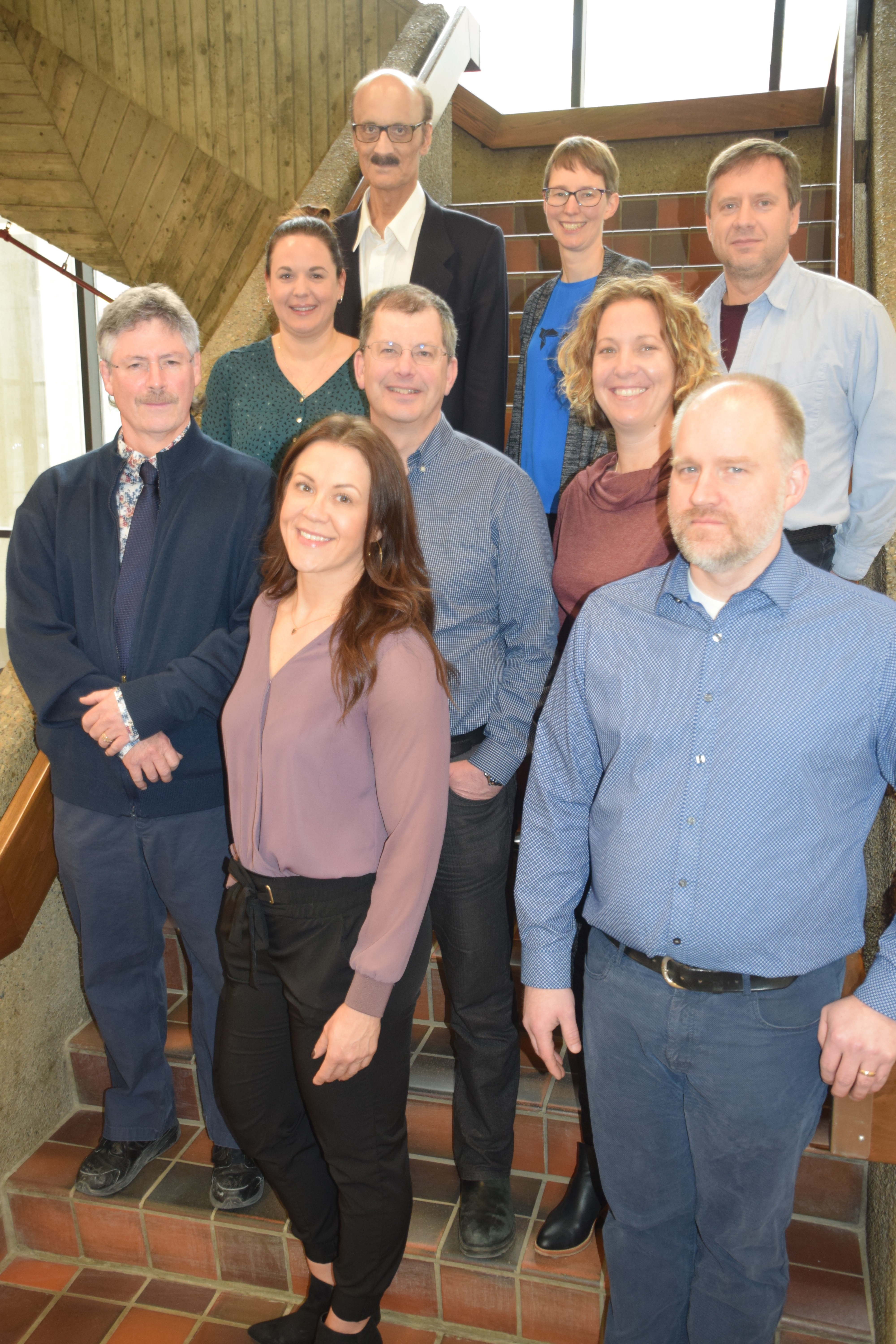
The Division of Anatomy Research Team: Back: Anil Walji, Jennifer Hocking, Hugh Barrett Middle: Karyne Rabey, Pierre Lemelin, Christine Webber Front: Daniel Livy, Kalyn McIntyre, Jason Papirny
The Division of Anatomy's researchers investigate basic science and clinical, including nerve, muscle and bone regeneration, retinal development and disease, gait and locomotor biomechanics, and cadaveric studies: hydrocephalus and nerve blocks, surgical, anatomical, orthopedic, musculoskeletal, histological, and neuroanatomical.
Dr. Daniel Livy is the Chair of the Division of Anatomy and primary contact for the instruction for the undergraduate and postgraduate gross anatomy. He is also the primary contact for those seeking access to the gross anatomy laboratory for either research or educational purposes. Research opportunities (surgical, anatomical, orthopedic, musculoskeletal, histological, neuroanatomical) are available in the gross anatomy lab for the Department of Surgery. Further, Dr. Livy continues to petition for a fresh-frozen cadaver facility at the University of Alberta, which will provide new research opportunities.
Dr. Pierre Lemelin is the supervisor of the Gross Anatomy laboratory and his personal research laboratory studies the convergence of gait and locomotor biomechanics among arboreal mammals, as well as the functional morphology and evolution of the primate hand. He co-edited The Evolution of the Primate Hand -Springer Publishing.
Dr. Jennifer Hocking leads a research laboratory studying retinal development and disease using zebrafish as a model system. Vision begins with light detection by photoreceptors and her team uses genetic manipulation, advanced microscopy, and electroretinography to study photoreceptor morphogenesis, function, and maintenance. Dr. Hocking is the primary embryology instructor within the Division of Anatomy.
Dr. Karyne Rabey’s research consists of interdisciplinary innovations to better understand the mechanics of movement (gait analysis) and exercise with musculoskeletal variation, sex differences and the mechanisms through which these are shaped. She utilises force plate mechanography, motion capture analyses, deep machine learning, micro-CT, and histology. Her lab directly tests relationships between bony morphology with behaviour, nerve injury, and muscle function.
Dr. Christine Webber’s laboratory investigates nerve regeneration and functional recovery using rodent and human models. Her work has shown that conditioning electrical stimulation prior to rodent nerve repair surgery promotes nerve regeneration in acute and chronic injuries, grafts, and nerve transfers. Other projects include neuroma formation and treatment, and investigations into axonal subtypes and their intrinsic rates of regeneration.
Dr. Anil Walji has had several cadaver research projects including investigations into skull diploic venous space as a potential route for cerebrospinal fluid diversion and absorption in the treatment of hydrocephalus, the use of ultrasound in cadaver-based trunk and peripheral nerve blocks. He is the author of several textbooks including a core anatomy undergraduate medical education textbook, Interactive Clinical Anatomy: A Workbook of Lecture Notes, Illustrations and Drawings.
Kalyn McIntyre is the Division of Anatomy administrator. She is the primary point of contact for the Division of Anatomy both internally (Department of Surgery and Faculty of Medicine and Dentistry) and externally (public relations). Ms. McIntyre advises undergraduate research students and medical elective students. She helps set up and manage our grants and she is our human resources coordinator.
Jason Papirny is the Program Coordinator of the Anatomical Gifts Program for our body donation program. Mr. Papirny supervises all access to the gross anatomy facility and each year; he organizes the Commemorative Service for the family of our donors.
Hugh Barrett is a laboratory technician in the Gross Anatomy lab and principal embalmer for the Anatomical Gifts Program. He helps facilitates all lab sessions by setting up and organizing specimens for the many different programs visiting our cadaver lab each day.