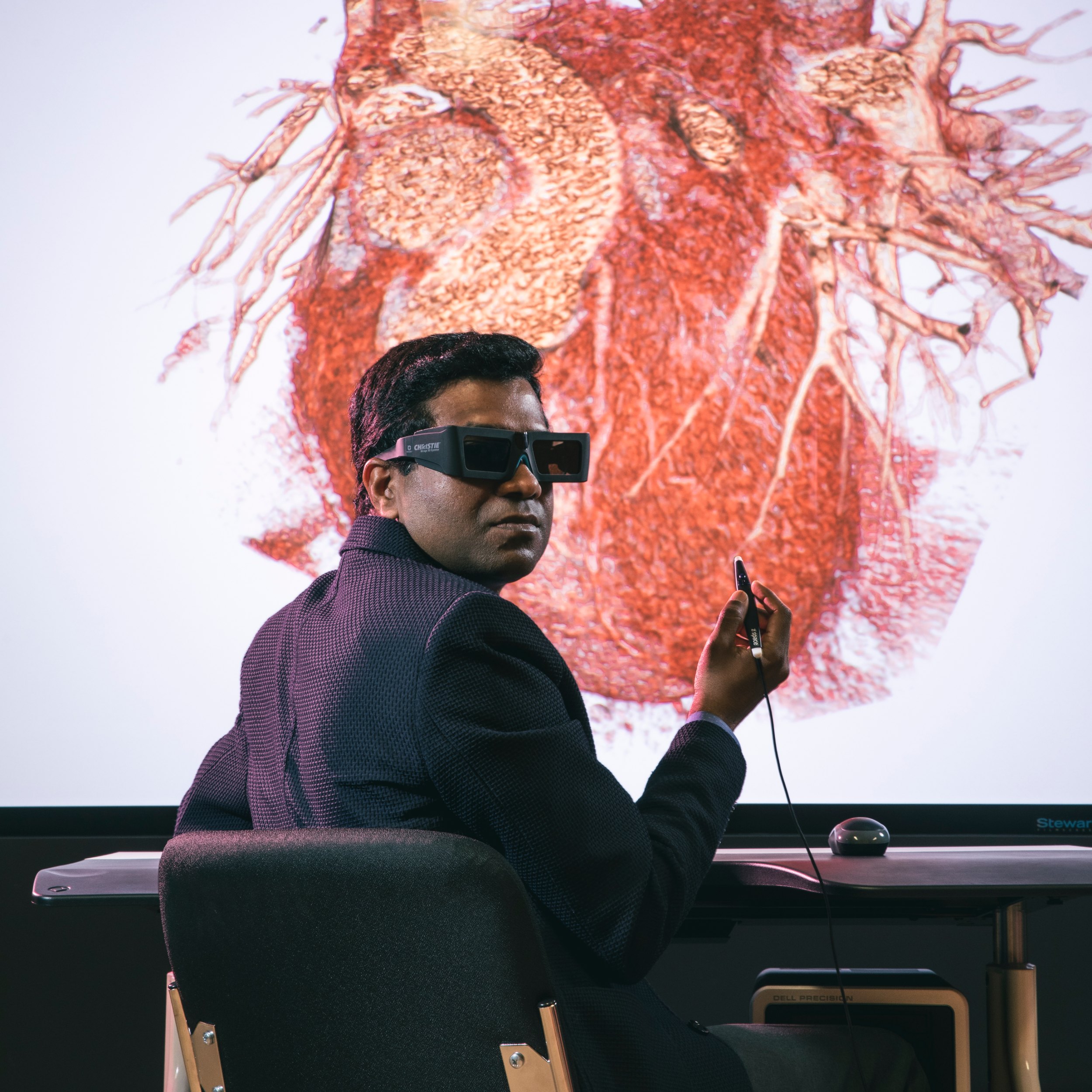
A University of Alberta-based team is developing a system to improve the way heart conditions are diagnosed by blending robotics and artificial intelligence with existing cardiac ultrasound technology.
“Current echocardiography (heart ultrasound) is used for virtually all patients with cardiac symptoms but it has limitations. For example, it doesn’t typically capture the entire heart in one single scan,” said Kumaradevan Punithakumar, associate professor (research) of radiology and diagnostic imaging in the Faculty of Medicine & Dentistry.
“We want to make the process more streamlined, so instead of looking at multiple scans in different windows, we have the entire heart in a single display,” said Punithakumar, who is also operational and computational director of the Servier Virtual Cardiac Centre, an advanced visualization lab for cardiac imaging at the Mazankowski Alberta Heart Institute.
The new system, which is being refined and developed for commercialization with new funding from Alberta Innovates’ Accelerating Innovations into CarE (AICE), would allow more patients to get scans and receive treatment more quickly.
“Digital health technologies are bringing the future to benefit patients today,” said Sunil Rajput, senior business partner for health innovation with Alberta Innovates. “The research by Dr. Punithakumar and co-workers is an excellent example, to rapidly collect heart images for clinicians while minimizing ergonomic stress on technicians."
“Our aim is to reduce waiting times and improve access to accurate and reliable imaging,” said Harald Becher, professor of medicine in the Division of Cardiology.
Precision diagnostics
In conventional echocardiography, a sonographer moves a probe around the patient’s chest to capture multiple 2D images of the heart from different angles, which are then individually examined by a physician to diagnose disease. Recently the technology has advanced so that multiple images can be captured in three dimensions, but they require the patient to stay very still for a long time by holding their breath, and each image is taken from only one angle so parts of the heart may be missed.
The U of A system has an optical tracking system that uses three cameras and markers attached to the ultrasound device to take multiple images from different angles during several breath holds. Then a software algorithm aligns and fuses the images, combining overlapping information and creating an accurate 3D picture.
In a recently published study, the researchers used the Multi-view Three-Dimensional Fusion Echocardiography System to measure the function of the left ventricle of 12 heart failure patients with pacemakers. The images were found to be as good as or better than those produced by standard echocardiography and by an advanced technique that requires the injection of a contrast agent to get better images.
“With the new method we could get comparable results without injection of a contrast agent and without taking the (small) risk of an allergic reaction,” said Becher. “We are confident that the new method will reduce the risk of errors and the need for additional imaging with more complicated methods like cardiac MRI (magnetic resonance imaging) or cardiac CT (computed tomography).”
Saving time, preventing injuries
The new system takes about 30 per cent less time per scan than traditional echocardiography, which could lead to shorter wait lists.
“Patients can wait as long as three months for electrocardiography,” Becher explained. “That’s a long time if you think you have a heart problem and you want to have it sorted out very soon.
Every week waiting for an ultrasound scan is lost time when it comes to starting treatment.”
The use of a robotic arm helps to prevent shoulder injuries, which are common for sonographers who perform echocardiography scans, Punithakumar added.

Becher predicted ultrasound will be the diagnostic technology of the future for cardiac patients.
“Ultrasound is not invasive, so it doesn’t hurt. And now, with our three-dimensional fusion system, we can get reliable results using less energy, less space and less time than with other technologies.”
Punithakumar and Becher noted their project was made possible by the U of A’s cross-disciplinary strengths in machine learning, engineering and cardiac clinical research. The development of the system is funded by Alberta Innovates, a Canadian Institutes of Health Research/Natural Sciences and Engineering Research Council of Canada Collaborative Health Research Project grant, the Canada Foundation for Innovation, Servier Canada and the Heart and Stroke Foundation. The team also has industrial partnerships with Siemens Canada and Medo.ai. Punithakumar is a member of the Women and Children’s Health Research Institute.
The researchers hope to have their system ready for broader clinical use within five years. First they will continue testing it on model hearts and with volunteers, and refining the software to ensure the highest accuracy.
Accelerating health innovation
This is one of six U of A projects that recently received funding from Alberta Innovates’ Accelerating Innovations into CarE (AICE) suite of programs, which support innovators to commercialize their health technology. The other projects are:
- “Mediation of gut microbiomes with ghost phages as alternatives to antibiotics,” Dominic Sauvageau, Faculty of Engineering
- “Personalized cancer vaccines using the Fusogenix genetic medicines platform,” John Lewis, Frank and Carla Sojonky Chair in Prostate Cancer Research funded by the Alberta Cancer Foundation, Faculty of Medicine & Dentistry
- “Next-generation ultrafast 3D ultrasound array technology for automated cardiovascular screening,” Roger Zemp, Faculty of Engineering
- “A multi-featured mobile application to support workflow of health-care aides who provide services to Albertans living with dementia,” Clinisys EMR and University of Alberta
- “Evaluating a new device for hand therapy—The FEPSim feasibility and usability study,” Karma Machining & Manufacturing and University of Alberta
