Galleries

Have a look at some image samples our faculty has captured using the scanning electron microscopes (SEM), transmission electron microscopes (TEM), and light microscopes (LM) in the Advance Microscopy Facility (AMF).

Click to enlarge the images in the three galleries.

Scanning Electron Microscope (SEM) Images

a cross section of a bassoon reed taken with a scanning electron microscope

Bassoon Reed (Long Section)

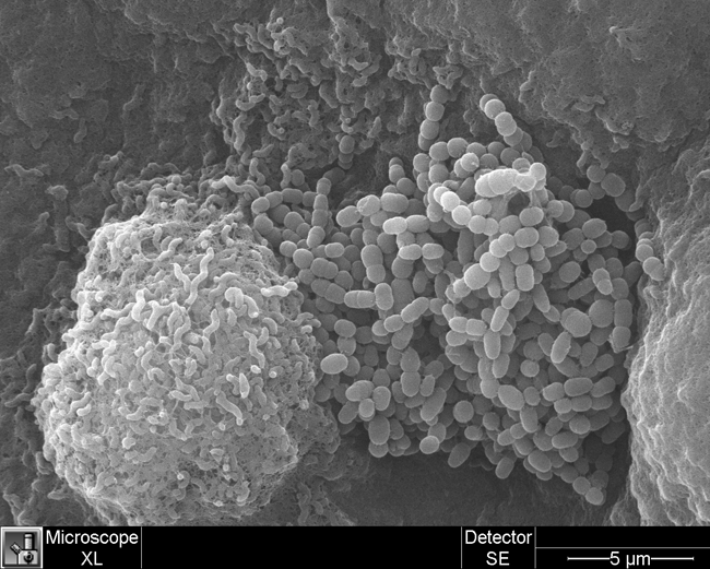
3D image of campylobacter jejuni and unidentified microbes, taken with a scanning electron microscope

Campylobacter Jejuni (on left) with Unidentified Microbes (on right)

a high magnification image of ciliated respiratory epithelium in lungs, taken with a scanning electron microscope

Ciliated Respiratory Epithelium (high mag)

a high magnification image of lung cells (ciliated respiratory epithelium), taken with a scanning electron microscope

Ciliated Respiratory Epithelium, 2 (high mag)

a low-magnification image of cliliated respiratory epithelium in lungs, taken with a scanning electron microscope

Ciliated Respiratory Epithelium (low mag)

the surface of a fish embryo, taken with a scanning electron microscope

Fish Embryo Surface

the surface of a fish gill, taken with a scanning electron microscope

Fish Gill Surface

a cross section of a leaf, taken with a scanning electron microscope

Leaf Cross Section

the surface of a leaf with visible trichromes, taken with a scanning electron microscope

Leaf Surface with Trichomes

3D image of a mouse artery wall with wavy elastin and red blood cells, taken with a scanning electron microscope

Mouse Artery Wall Showing Wavy Elastin and Red Blood Cells

3D image of a plant filament, taken with a scanning electron microscope

Plant Filament

3D image of pollen grains, taken with a scanning electron microscope

Pollen Grains

A low magnification image of a sequoia leaf, taken with a scanning electron microscope

Sequoia Leaf (low mag)

Transmission Electron Microscope (TEM) Images

a cross section of bacteria cells, taken with a transmission electron microscope

Bacteria in Cross Section

a negative stain of bacteria cells with flagella, taken with a transmission electron microscope

Bacteria with Flagella (Negative Stain)

a negative stain of bacteria cells with pili, taken with a transmission electron microscope

Bacteria with Pili (Negative Stain)

Three bacterophages, taken with a transmission electron microscope

Bacteriophage

a muscle capillary with endolithia and red blood cells, taken with a transmission electron microscope

Capillary within Muscle, Showing Endothelia and Red Blood Cells

collagen fibers, taken with a transmission electron microscope

Collagen Fibers

connective tissue in a mouse kidney, taken with a transmission electron microscope

Connective Tissue in Mouse Kidney

microscopic image of muscle fibers and mitochondria, taken with a transmission electron microscope

Muscle Fibers and Mitochondria

microscopic image of a pacific hagfish's liver cells, taken with a transmission electron microscope

Pacific Hagfish Liver Cells

photoreceptor cells of a mouse retina, taken with a transmission electron microscope

Photoreceptor Cells of Mouse Retina

microscopic image of a plant cell with 7 large starch granules, taken with a transmission electron microscope

Plant Cell with Large Starch Granules

microscopic image of a plant cell with visible organelles and a thick cell wall, taken with a transmission electron microscope

Plant Cell with Organelles and Thick Cell Wall

a microscopic image of the plastids within a plant cell, taken with a transmission electron microscope

Plastids within a Plant Cell

a microscopic image of silver nanoparticles, taken with a transmission electron microscope

Silver Nanoparticles

a microscopic image of a virus shaped like two side-by-side circles, taken with a transmission electron microscope

Virus (Negative Stain)

Light Microscope (LM) Images

a microscopic image of a cross section of an amphibian's toe, taken with a light microscope

Amphibian Toe Cross Section

a microscopic image of a clam's foot, taken with a light microscope

Clam Foot (Masson's)

a microscopic image of an fluorescent elm cross section, taken with a light microscope

Elm (Fluorescence)

a microscopic cross section of a fish using fluorescence, taken with a light microscope

Fish (Fluorescence)

a microscopic image of a cross section of lamprey cells, taken with a light microscope

Lamprey (H&E)

a microscopic image of a cross section of the cells in a mouse duodenum, taken with a light microscope

Mouse Duodenum (PAS)

a cross section of peony cells, taken with a light microscope

Peony (Safranin O & Fast Green)

a microscopic image of polychaete in callyspongia. taken with a light microscope

Polychaete in Callyspongia (Masson's)

a cross section of cells in a plant seed, taken with a light microscope

Seed (Toluidine Blue)

a microscopic image of a dissection of a spruce cone, taken with a light microscope

Spruce (Dissecting Scope)

a cross section of spruce cells, taken with a light microscope

Spruce (PAS)

a microscopic image of spruce cells, taken with a light microscope

Spruce (Toluidine Blue)

a cross section of tadpole snail cells, taken with a light microscope

Tadpole Snail (Masson's)

a cross section of western red cedar cells, taken with a light microscope

Western Red Cedar (Safranin O & Fast Green)

a microscopic cross section of wood cells, taken with a light microscope

Wood (Toluidine Blue - DIC)